20년도 OX 모음
- The (cathode) is the part of the X-ray tube where (negatively) charged and accelerated (electron-cloud) move to target more intensively after kV is applied to the tube.
- Reduction of the X-ray intensity at the (anode) side of an X-ray field it can be observed that the film density is lower on the (anode) side. This phenomenon is termed the (Heel effects).
- (Grid cutoff) is meaning term for differences in radiographic intensity that are caused by improper focusing of the lead lines of a grid.
- (Fluoroscopy) is an imaging technique commonly used by physicians to obtain real-time moving images of the internal structures of a patient through the use of an X-ray source.
- (Intensifying screen) is a kind of device consisting of fluorescent material which is placed in contact with the film in a radiographic cassette. Radiation interacts with the fluorescent (phosphorus), releasing light photons. These photons expose the film with (greater) efficiency than would the radiation alone. Thus, patient exposure to radiation can be (reduced).
- (Collimator) is a device for limiting the size and shape of a radiation beam. It is used to reduce (scattered radiation). decrease the patient dose, and increase radiographic quality.
영상 진단에 사용하는 장비 | 구현에 사용되는 에너지원 | 영상의 밝고 어두움을 나타내는단어
- Radiography | X-ray, Gamma-ray | Radiopaque/Radiolucent
- CT | X-ray | Hyperattenuation/Hypoattenuation
- MRI | RF(radiofrequency) 에너지와 강한 자기장) | Hyperintense/Hypointense
- Ultrasonography | 초음파 | Hyperechoic/Hypoechoic
교통사고로 척추손상이 의심되는 환자가 내원했을 때 사용할 영상진단 장비 3종
- X-Ray : fracture check, 방사선 촬영은 척추의 골절 또는 탈구를 신속하게 평가하는 데 유용
- CT : Surgery plan, 복잡한 골절이나 탈구의 평가에서 유용, 빠른 획득 속도로 3차원 영상을 제공하며, 척추 골절의 위치와 형태를 명확히 파악할 수 있어 수술 계획에 도움
- MRI : Soft tissue damage check, CT나 방사선 촬영으로는 확인하기 어려운 척수의 압박 정도와 soft tissue 손상 부위를 정확하게 파악
X-ray machine is also known as ( A ). Radiographers can adjust ( B ) and ( C ). ( D ) has ( E ) on tungsten filament. ( F ) shot positively charged ( H ). X-ray is converted 99% of ( H ) and 1% of ( I ).
- X-ray machine은 다른 말로 A) radiography라고도 함.
- B) mAs와 C) kVp에 의해 조절됨.
- mAs : 전자무리의 양.
- kVp : 충돌량의 차이를 만들어냄.
- D) Cathode는 필라멘트에서 E) electron cloud(전자무리)를 만듦.
- F) Electron이 관을 지나가서 H) anode인 target(tungsten plate)에 충돌함.
- 충돌 결과 이온화되어 99%는 H) heat, 1%는 빛과 I) X-ray로 발생.
The ( ) is part of an x-ray tube and serves to expel the ( ) from the circuit and focus them in a beam on the focal spot of the ( ). It is a controlled source of electrons for the generation of x-ray beams. The electrons are produced by heating the filament i.e. a coil of wire made from ( ), placed within a cup-shaped structure, a highly polished nickel focusing cup, providing electrostatic focusing of the beam on the anode.
- Cathode, Electrons, Anode, Tungsten
X-ray 영상을 개선할 수 있는 테크닉들과 그 이유 (Limitation of Scattered Radiation)
- 소아과나 움직임이 심한 환자의 경우, 조사 시간을 줄이고 그에 맞추어 mA를 높인다.
- Collimator : 불필요한 방사선을 막고, scattering 줄여주며, 조사 중점을 표시해줌.
- Reduction of tissue amount : 두꺼운 몸체를 가진 대형견의 경우, 복부를 눌러 두께를 줄여 촬영하면 scattering을 줄일 수 있음.
- Prevention of back scatter : 납으로 된 cassette back을 테이블 뒤쪽에 깔아, 산란선이 다시 튀어서 필름에 scattering을 만드는 일이 없도록 방지.
- Grid : 적절한 grid를 사용하면 scattering을 줄일 수 있음.
Grid의 유용성은?
- 산란선은 1차선과 다른 방향으로 다시 필름지로 들어가는데, 이 산란선을 제거해주는 체 역할을 한다. 알루미늄이 들어간 납선으로 만들어 깨끗한 이미지로 개선시켜준다.
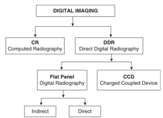
Digital radiography (=DR)의 종류와 특징
- CR(Computed Radiography) : 필름에 전기 저항값을 측정하는 실리콘 detector가 있어, X-ray 신호를 전기적 신호로 변환.
- DDR (Direct digital radiagraphy)
- CCD (Charged coupled device) : 미세한 카메라들이 영상을 읽음.
- Flat Panel (Digital Radiography)
- Indirect : 증감지에서 나온 빛을 아래의 전자 센서가 받아 영상화함.
- Direct : 증감지 대신 selenium photoconductor가 위치하고, silicone detector가 빛의 세기를 인식해 영상화함.
Radiographic quality
Geometic unsharpness - X-ray에서 해부학적 구조물이 실제 모양/크기와 달라지는 경우
- focal spot size가 클 때 : 반음영 효과(penumbra effect)
- decreased SID : X-ray 발생원과 검체 사이가 가까울 때 블러 현상 발생
- Motion : 촬영하는 동안 움직이면 경계선이 불명확해 보임, elongation과 foreshortening 발생
- Certain screen / film combination : 서로 다른 회사 제품을 사용했을 때
- X-ray 발생원에서 멀리 떨어져 촬영하면 elongation 또는 foreshortening 발생
- 검체와 수평이 맞지 않게 촬영하거나, 조사 중심으로부터 멀게 촬영
Geometric distortion과, magnification을 줄이는 방법을 서술하시오.
- X-ray 발생원에서 멀리 떨어지거나 검체와 수평이 맞지 않는 등 보정을 잘못하면 짧아보이거나(foreshortening), 확대되어 보일 수 있음(elongation).
- 따라서 바닥에 최대한 가깝게 붙여서, 조사 중심을 맞추어 촬영해야 한다.
kVp를 높여야 하는 환자 상태를 열거하고 그 이유를 쓰시오.
- 매질의 두께가 두꺼워지는 경우, 검체의 density가 높은 경우 kVp를 증가시켜 투과도를 올린다.
- Pleural effusion (흉수)
- Pulmonary fluid (폐에 액체 고임)
- Pneumoconiosis (진폐증; 폐에 먼지 축적)
- Pulmonary fibrosis (폐섬유화; 노령에서)
- Bronchiectasis (기관지확장증)
- Post-surgical thoracic study (수술후 흉부검사)
- Cardiomegaly (심장비대)
- Pericardial fluid (심막의 액체)
- Ascites (복수)
- Large abdominal mass (복부 종괴)
- Ingesta filled GIT (내용물로 가득찬 위장관)
- Soft tissue edema & calcification (연조직 부종 & 석회화)
kVp를 낮춰야 하는 환자 상태
- 매질의 두께가 얇아지는 경우, 검체 density가 낮은 경우 투과도를 낮춘다.
- Focal destructive bone disease (국소성 파괴성 골질환)
- Generalized bone disease (전신성 골질환)
- Emphysematous soft tissues (기종성 연조직; 연조직에 공기 포함)
- Pneumothorax (기흉; 흉강 내 공기 찬 상태)
- Air filled megaesophagus (공기로 찬 거대식도)
- Massive pulmonary air trapping (폐에서 공기가 빠져나가지 못함)
- Aerophagia with dilated air filled stomach (공기삼킴증 → 위가 공기로 팽창)
- Gastric dilation/torsion syndrome (위 팽창/염전 증후군)
- 등..
Basic radiographic opacity
- Radiopaque = 불투과성 = more white
Radiolucent = 투과성 = more black
- opaque : 물질의 원자번호가 클수록, 조직의 두께가 두꺼울수록, 같은 매질이 중복될 때.
- 원자번호 : gas < fat < water / soft tissue / muscle < bone < metal
- Bone opacity : compact bone, trabecular bone, cortex가 더 밝다(lucent).